Flow cytometry platform
Our Research Unit has massively invested to setup a flow cytometry facility, which comprises :
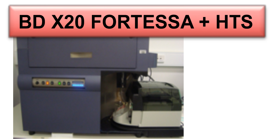
BD FORTESSA X20
(acquired in 2018)
4 lasers, 18 colors
X20 HELP PAGE. Link to booking.
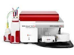
BD ACCURI C6
(acquired in 2015)
2 lasers, 4 colors
C6 HELP PAGE. Link to booking.
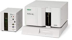
BioPlex Luminex (BioRad)
(acquired in 2013)
2 lasers, 3 colors, High-Throughput System for multiplex cytometric bead arrays
Link to booking.
FACSARIA III cell sorter (BD Biosciences)
(acquired in 2010)
5 lasers, 15 colors, 4 way sorting on plates, slides or tubes (acquisition for the Bichat site with grants obtained from the CODDIM, INSERM and the Université Denis Diderot).
ARIAIII HELP PAGE. Link to booking.
These devices allow to perform analytical flow cytometry on vascular or hematopoeitic cells or microparticles. We have an extended expertise for multiplexed measurements of analytes with cytometric bead arrays (CBA, Luminex, eBiosciences and customized beads). The new cell sorter provides us with phenotypically pure cell preparations for further cell cultures or molecular biology.
Currently, we do not have a dedicated core lab engineer and our strategy is to promote internal trainings of our users (2 training sessions per year are organized by A Nicoletti). We now have a consistent number of skilled users that can in turn train the beginners.
Live cell imaging and analysis
Our research Unit acquired instruments for live cell functions analysis:
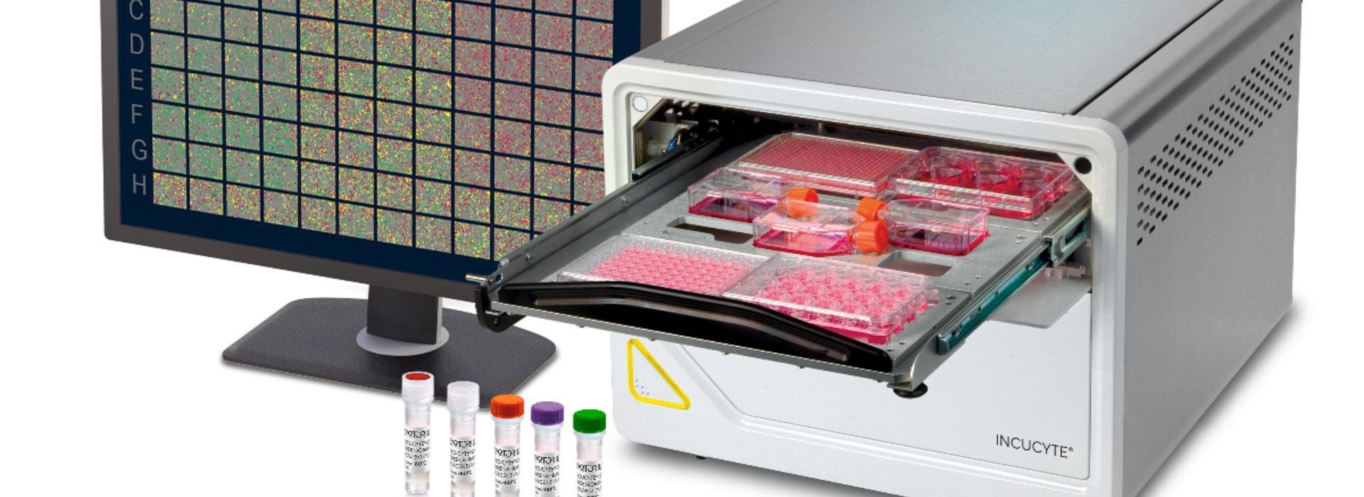
Incucyte® SX5 Live cell Imaging System allows to acquire and view images over time (Fluorescence and HD phase imaging modes, Minimize cell disturbance with a mobile optical train).
XCelligence Agilent uses live, simultaneous, and real time biosensor impedance-based and image-based measurements.

In addition, we acquired the Lonza’s 4D-Nucleofector System, a modular system for the efficient transfection of primary cells and cell lines with a variety of substrates including plasmid DNA and siRNA.

Coagulation analysis and platelet function
Our research Unit has invested in technologies allowing coagulation analysis and characterization of human and mouse platelet functions.
Coagulation analysis
ROTEM®delta
The ROTEM delta is an integrated all-in-one system allowing the measurement of thromboelastometry parameters in platelet poor plasma or whole blood. This viscoelastometric method measures the interactions of coagulation factors, inhibitors and cellular components during the phases of clotting and subsequent lysis over time during a one-hour time reaction at 37°C. This system can assess a series of coagulation parameters such as clotting time (CT), clot formation time and firmness (CFT and MCF) and also fibrinolytic parameters as maximal lysis (ML) and lysis time and rate (LT, CLR).

StarMax
The coagulation analyzer StartMax is a semi-automated benchtop analyzer for all clotting assays from routine to specialized assays. Measurements can be qualitative or quantitative using small volumes of human or mouse materials.

Platelet function
The “platelet platform” has many applications including:
- Basic research studies on the physiologic role of platelets in hemostasis and their pathologic role in thrombosis
- Identification of platelet defects in patients
- Clinical studies: diagnostic markers and platelet responsiveness to therapeutic agents
- Bioengineering: analysis of the biocompatibility, hemostatic toxicity of polymers and devices; development of new tracers.
The platform can acquire the following parameters:
- Hematological parameters can be analyzed by a human or mouse blood analyzer. Micro-sampling from whole blood allows real cell volume measurement:
– Sysmex XE for platelet quantification and volume
– ABX Pentra60 Horiba for Human hemocytometry
– SCIL Vet ABC for mouse hemocytometry


- In vitro bleeding time is acquired with a PFA100 Siemens
Platelet phenotype is analyzed by flow cytometry (see above), immunoblot and immunoprecipitation. - Platelet activation is assessed by analyzing expression of activation markers by flow cytometry
Platelet aggregation is measured by light aggregometry (8 channels aggregometer: APACT 40004, Elitech)

- Platelet procoagulant activity is assessed by thrombinography on platelet rich plasma. It consists in measuring the catalytic activity of platelets on the formation of thrombin.

- Platelet adhesion in static conditions is performed by colorimetric analysis.
- Shear-induced platelet activation is analyzed with “Couette” devices (Hospital hematology laboratory). The principle is to subject platelets to increasing shear rates in order to reproduce the conditions of a stenosed vessel. Platelet activation is measured as the disappearance of individual platelets.

In vitro cell adhesion, thrombosis and thrombolysis can be studied under flow Microfluidic flow chambers. The laboratory has the possibility to analyze the capacity of platelets to form thrombi in whole blood under flow condition with platelets being labeled with a fluorescent probe (DiOC6 or rhodamine). Two types of flow chambers are available: the classical Maastricht chamber and the small volume dispensable chambers from Cellix (Dublin, Ireland) either coated with purified proteins or covered by an endothelial cell monolayer. Modulation of flow conditions is possible with the use of Exigo and/or Myrus pumps and offer the possibility to work at defined shear stress.
A specific protocol developed by LVTS T6 allows real time analysis of clot formation and lysis in human whole blood under arterial or venous flow rates and offers a way to evaluate the lytic efficacy of drugs during thrombolysis.


In vivo thrombosis is studied using the ferric chloride model, platelets being labeled in vivo by the injection of rhodamine 6G. Arterioles and venules are visualized with an inverted epifluorescent microscope (×10) (Nikon Eclipse TE2000-U), coupled to Metamorph7.0r1 software (Universal Imaging Corporation). Platelet deposition and thrombus growth in arterioles and venules are monitored in real-time until complete occlusion occurs. The analyzed parameters are the vessel occlusion time and the surface and volume of platelet aggregates.
Platelet adhesion is monitored in real time using fluorescence microscopy with a monochromator epifluorescence illumination system (DMRIB Leica) and a DP30W camera (Olympus).
In vivo fibrinolysis is analyzed through a new and original method that we have developed: ferric chloride thrombosis is triggered on vessels exposed on a dorsal skinfold chamber. These methods allow studying clot lysis and its pharmacological regulation by intravital microscopy on animals maintained alive.
Biochemical analysis
We have several plate readers including two recently acquired:
Promega GloMax Explorer System: advanced detection capabilities (Absorbance, Fluorescence, Luminescence with Dual injector).

Thermo Scientific™ Varioskan™ LUX: end point or kinetic readings of fluorescence, absorbance or luminescence.

We also acquired materials for different analysis:
Bench top analyzer: Diasys Respons®920 bench top analyzer can titrate various analytes in any human or animal fluid sample.

Automated capillary-based western blotting system: Protein Simple WES (R&D sytems) combines capillary electrophoresis and immunodetection and allows multiplex analyses.

Thermo Scientific™ iBright 1500 The iBright FL1500 Imaging System supports major imaging applications, fluorescently stained protein gels, colorimetric stained protein gels, and colorimetric membrane dyes for membrane-based western blotting.

Genomic platform
The Genomic platform (headed by Pr C. Boileau, T2 and in the future by C. Le Goff and Pauline Arnaud) houses the platform which has the equipment to perform state-of-the-art medium throughput genetic studies (DNA and RNA panel and whole exome sequencing). COFRAC ISO 15189 certification is underway in the Department.
Since its creation, the platform equipment has been acquired through combined hospital, University and INSERM funding. Presently there are 3 NGS machines (Ion S5 XL System from System Thermo Fisher Scientific; MiSeq and NextSeq 500 from Illumina) and automation for library preparation, 2 x 3500XL (Applied Biosystems) Sanger sequencers, quantitative and droplet PCR machines. The latest NextSeq 500 machine was co-funded by INSERM and Plan Maladies Rares. The major current technological developments concern new approaches in RNA sequencing and, depending on funding, third generation sequencing and ‘long read sequencing’. All equipment is maintained by dedicated hospital lab personnel and biologists. The data generated by the platform are stored on a dedicated server and the current pipelines are housed in hardware located at INSERM U1137 (IAME for “Infection, Antimicrobials, Modelling, Evolution”, located on site in the Medical School Building). The material was obtained through a grant (“Actions Structurantes”, CATIBiomed) from Université Paris Cité and added co-funding from the 5 INSERM research Units located on site. Finally, bioinformatic scientist (Plan Maladies Rares funding) is dedicated to raw data analysis while dedicated IAME personnel is in charge of hardware.
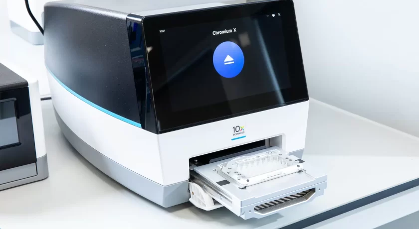
Our last equipment is the 10X-Chromium for high-throughput single-cell RNA sequencing (scRNA-seq). It enables the analysis of gene expression profiles at single-cell resolution. Also, it can operate at a scale of hundreds to ten thousands of cells per run. 10x Chromium is compatible with the droplet-based scRNA-seq methods of 10x Genomics.
PhysicoChemical platform
In addition to dedicated rooms for chemistry (inorganic, organic and polymer), our research Unit provides to its members its own equipment allowing the physico-chemical characterization of polymers and colloids such as:
The determination of polymer properties: fusion & crystallization events, glass transition, chemical functions, composition, absolute molar mass, refractive index, dn/dc values, contact angle, viscosity, … can be achieved with Differential Scanning Calorimetry (DSC), Fournier Transform Infra-Red (FT-IR), UV and fluorescent spectrometers, Nuclear Magnetic Resonance 400 & 500 MHz (NMR-shared equipment USPN), MultiAngle Laser Light Scattering (MALLS – collaboration), Refractometer, Contact angle goniometer, Rheometer.
The determination of colloidal properties: second virial coefficient, translational diffusion coefficient, radius of gyration, hydrodynamic radius, polydispersity, surface charge, shape, two-dimensional image, visualization, … are achieved with Multi-Angle Laser Light Scattering (MALL, Dynamic Light Scattering (DLS), Laser Doppler Velocymetry, Laser diffraction particle size analysis, Atomic force Microscopy (AFM- shared equipment USPN), Field-Emission Gun Transmission Electron Microscope (FEG-TEM- shared equipment USPN), Scanning electron Microscopy (SEM – shared equipment USPN).
Physical properties of hybrid nanomaterials are carefully characterized allowing in-situ monitoring through magnetometry and various optical spectroscopies: total reflection X-ray fluorescence spectroscopy, Raman, FTIR, UV-visible, Near infrared transmission and luminescence.
S2 PICOFOX – Fast Trace Element Analysis with TXRF (Bruker)/spectrofluorimeter/ VersaLab™ 3 Tesla magnetometry measurement system.


The mechanical characterization of soft materials has been essentially developed during the last years with:
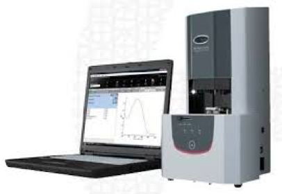
ElastoSens™ Bio is a smart, non-destructive and patented analytical instrument that unlocks the access to crucial information on the viscoelasticity of soft biomaterials and medical devices for R&D, quality insurance and process control.

Piuma nanoindenter is a powerful table-top instrument to explore the micro-mechanical properties of biomaterials and hydrogels down to cell-length scales.

Discovery Hybrid Rheometer-3 (TA Instruments) incorporates the latest technological advances from the world leader in rheology and. offers direct strain control, as well as unparalleled normal force measurement accuracy.


Plateform of Nanomaterial Characterization of the Paris 13 University
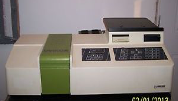
V-630 UV-VIS Spectrophotometer (Jasco) Kontron and Uvikon 941 Spectrophotometer
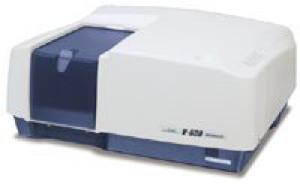
Available for analysis over a wide wavelength range of 190 to 1,100 nm, stray light of less than 0.04% and a spectral bandwidth of 1.5 nm.
Thermo Scientific Nicolet 380 FT-IR Spectrometer
The Nicolet 380 FT-IR spectrometer is available for analysis in the mid- IR regions. A real diamond ATR accessory can be easily installed.
Bruker Tensor 27 IR
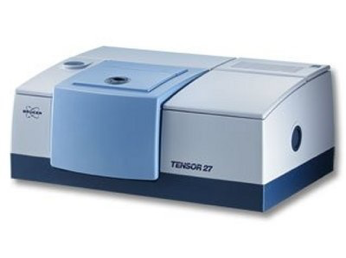
The Bruker Tensor 27 has several accessories that allow for a variety of sample measurements. Solids and liquids can be measure.
VersaLab™ 3 Tesla, Cryogen-free Physical Property Measurement System
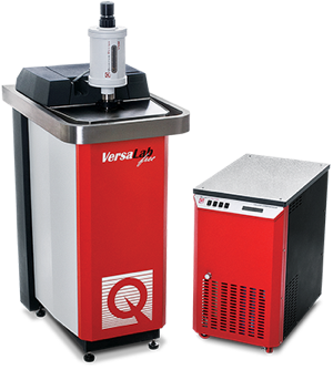
With a temperature range of 50 – 400K, this 3 tesla platform is perfect for accomplishing many types of materials characterization in a limited space.
Nano-ZS (Red Badge) ZEN 3600 device (Malvern Instruments, Malvern)
The Zetasizer Nano ZS is a high performance two angle particle and molecular size analyzer for the enhanced detection of aggregates and measurement of small or dilute samples, and samples at very low or high concentration using dynamic light scattering with ‘NIBS’ optics. The ZSP also incorporates a zeta potential analyzer that uses electrophoretic light scattering for particles, molecules and surfaces, and a molecular weight analyzer using static light scattering.
LabsSys evo TG-DTA-DSC 1600 device (Setaram)
TGA, and Simultaneous TGA-DTA, TGA-DSC from ambient to 1600°C.
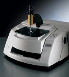
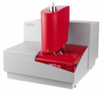
Spectrofluorometer Spex FluoroMax Jobin Yvon Horiba
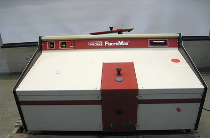
A spectrofluorometer used to obtain fluorescence excitation and emission spectra to quantify characteristics of a fluorophore-containing solution.
Spectral Range: 200 to 900 nm.
Hotte à poudre Poste de pesées sécurisées
Bobigny site equipment
Spectrofluorometer FluoroMax-4 Horiba
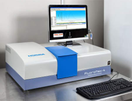 The FluoroMax is a compact spectrofluorometer which offers a great sensitivity in fluorescence investigations as well as features not found in most table-top fluorescence detection systems.
Contact : Laurence MOTTE-Yoann Lalatonne, Frederic.Geinguenaud
Microonde Monowave 400 – Anton Paar
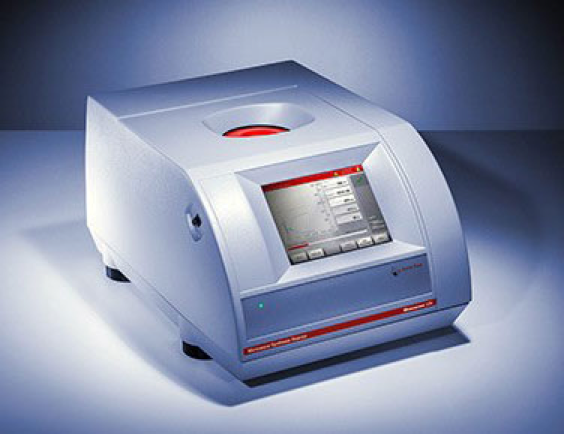 Monowave 400 – Anton Paar est un réacteur micro-ondes hautes performances spécialement conçu pour les applications de synthèse micro-ondes à petite échelle dans les laboratoires de recherche et de développement. De nos jours, le rayonnement micro-ondes n’est pas seulement utilisé avec succès pour l’analyse organique – la synthèse inorganique, la science des matériaux, la chimie des polymères ainsi que d’autres disciplines peuvent également être réalisées avec succès.
Contact : Laurence MOTTEYoann Lalatonne, Frederic.Geinguenaud
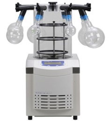
A lyophilizer Alpha 1-2 LDplus (CHRIST)
 Contact : Laurence MOTTEYoann Lalatonne, Frederic.Geinguenaud
S2 PICOFOX – Fast Trace Element Analysis with XRF (Bruker)
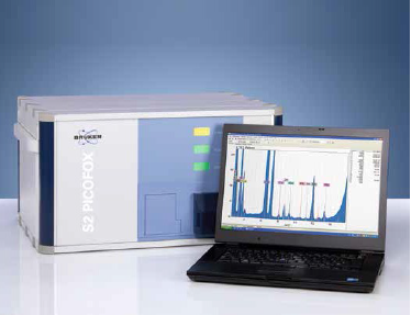 The S2 PICOFOX is the world’s first portable benchtop spectrometer for fast quantitative and semi-quantitative multielement microanalysis of liquids, suspensions, solids and contaminations using the principle of total reflection X-ray fluorescence spectroscopy. Reaching detection limits in the ppb and ppm range the S2 PICOFOX is optimally suited for trace element analysis.
Contact : Laurence MOTTEYoann Lalatonne, Frederic.Geinguenaud
Biacore X100 processing unit
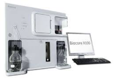 Real-time insights into protein function & biological mechanisms
Kinetics, affinity, specificity and concentration analysis in one system
Define structure/function relationships
Understand the dynamics of molecular pathways
In development and research studies, select promising molecules that could be novel targets for research use, diagnostics, or therapy.
Develop and run assays for interactions involving LMW compounds
Strong support from assay development to data interpretation
Contact: Haddad Oualid bureau 228 Tel: 01 48 38 76 97
BioSpec-nano
 Le BioSpec-nano est un spectrophotomètre approprié pour effectuer des contrôles de concentration d’ADN et d’ARN dans des échantillons d’acide nucléique. La quantification de l’ADN et de l’ARN peut être simplement et rapidement réalisée avec de très petits échantillons à partir du microlitre. La vitesse d’analyse est améliorée grâce à des fonctions dédiées, comme le montage automatisé de l’échantillon, et logiciel d’exploitation facile à utiliser.
Contact :Haddad Oualid –Angela Sutton
Applied Biosystems StepOne™
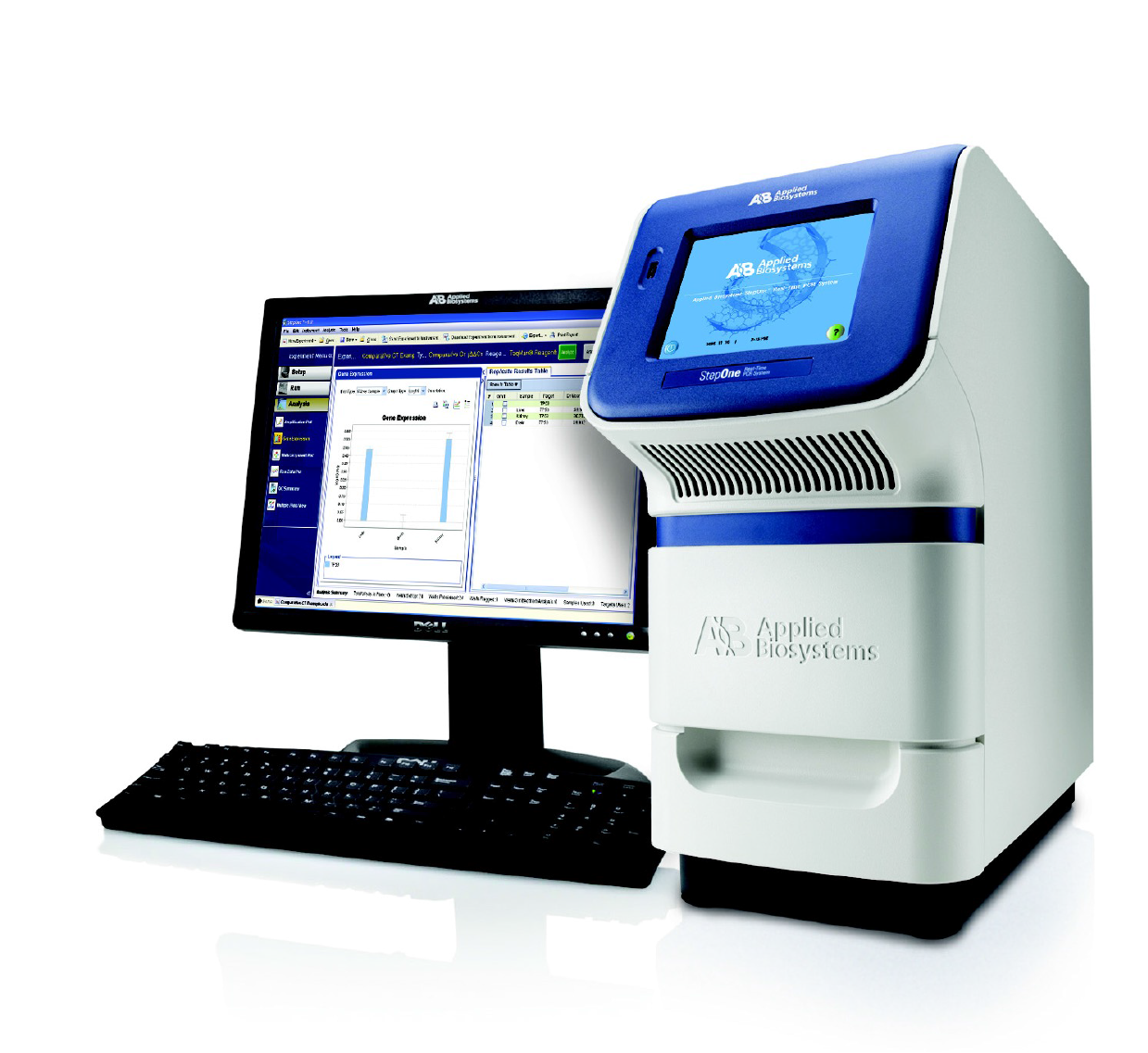 Système de PCR en temps réel
Expériences de quantification relative par la méthode des courbes standard et de comparaison des valeurs de CT.
Contact : Haddad Oualid –Angela Sutton
Immunohistochemistry facility
Paraffin embedding/sectioning
Leica ASP300 tissue processor for paraffin embedding
Thermo Fisher Microm MYR-EC350 (modular tissue embedding center, including dispensing console and cryo console with a large cooling surface)

Leica RM2245 and Microm HM340E microtomes

Resin embedding
Tissue embedding in special resins is required for transmission electron microscopy as well as for analysis of hard samples (stented vessels, calcified tissues). Samples should be cropped using a linear precision saw (Buehler ISOMET 5000) and polished (Buehler METASERV 250 grinder-polisher) before sectioning with a classical microtome equipped with a gem grade diamond knife (5-15 um). Ultra-thin sections are obtained using Reichert-Jung Ultracut-E, for electron microscopy analysis (performed at Bichat faculty of medicine).
Cryosectioning
Microm HM525 cryostat
Cryostar NX50 op cryostat

Observation
Several microscopes allow routine observation (Nikon Eclipse E400, Olympus BH-2…).
A high throughput slide scanner for brightfield and fluorescence (Nanozoomer 2.0 Hamamatsu) is available for the whole research Unit. A new slide scanner with higher capacity and performance (S60 Hamamatsu Nanozoomer) is being acquired for the platform since our project was selected for the 2022 Sesame call. The new slide scanner is part of the UMS 34 Histology platform and therefore opened to all unit of the site, to other academic research groups and accessible to industrial partners. Image files are stored on internal server and can be viewed via intranet. Software for analysis and quantification currently used are Histolab, Archimed and Cartograph (Microvision).

Inverted fluorescence microscopes Zeiss 6D microscope system and LEICA DMI8 with motorized stage and LED sources for fluorescence applications where rapid excitation changes are required allow image acquisition (including cartographies) of classical immunohistochemical and fluorescent preparations and the realization of time-lapse movies in flow chamber and mouse intravital microscopy experiments. The Zeiss microscope is also equipped with an apotome module for the acquisition of multi-channel optical sections.

Macroscopic observation and acquisition through 3 Leica MacroFluo: (1) Leica MacroFluo Z16 Apo A which is a fully apochromatic zoom system for high contrast, high-resolution, detailed analysis. Zoom, iris diaphragm and fine focus are motorized. The single beam path provides 2D images and ensures parallax-free. Images are delivered through an Andor’s DSD Revolution system which is an innovative hybrid of spinning disk technology and structured illumination, and are captured with an Orca Flash 4.0 video camera which is highly sensitive, displays a high dynamic range, is fast, has a large field of view, and excellent resolution. This equipment is particularly suited to follow in real time fluorescence in tissues on live animals (Acquired with an Ile de France CORDDIM grant). (2) Leica MacroFluo Z16 equipped with a Cool-SNAP camera and Leica MacroFluo Z16 equipped with a Jenoptic camera.

Confocal microscopy and bi-photon microscopy are performed on site at Bichat School of Medicine.
We have acquired a scanning electron microscope from JEOL (JSM-IT100LA) installed in June 2017 (Ile de France CORDDIM grant).
Functional investigation
Restricted access to LVTS’s members. You should be logged in to see the content
Preclinical imaging platform
The platform equipment for in vivo imaging of small mammals includes ([iso] means iso9001:2000 certification):
In vivo functional explorations
- Non-invasive blood pressure measurement: Tail-cuff device for mice and rat: Phymep and AD Instruments.
- Metabolic cages for mice: Phymep and Marty technology.
- Electrocardiography Surface recording and analysis: Emka IOX 1.8 and ECG-Auto software 1.5.12.36.
In vivo and ex vivo imaging
Doppler echography imaging device
Vevo 3100 (Visualsonics; acquired Dec 2022, with VevoStrain Analysis Suite, AutoLV Analysis Software, 55 MHz MX Series transducer) and Vevo 2100, with 9-18 MHz and 22-55 MHz probes and software for myocardial strain analysis (obtained in 2010).

SPECT imaging device
NanoSPECT/CT Multi-pinholes (Mediso-RS2D) with « Nucline » and « TeraTomo3D » SPECT software for mouse, rat and rabbit high resolution imaging (obtained in 2010).

PET-MRI imaging device
NanoPET-MRI 1 Tesla (Mediso-RS2D) with «TeraTomo3D» and Pmod softwares for mice and rats (obtained in 2017).
Body mouse and rat coils.

MRI imaging devices
- Spectrometer PharmaScan 70/16 imager 7 Tesla (Bruker) with ParaVision Package and holders for rats and mice (obtained in 2010)
- Spectrometer BioSpec 70/30 imager 7 Tesla (Bruker) with ParaVision Package and holders for rats and mice (obtained in 2015).
Body mouse and rat coils, head and heart rat coils, surface coils.


Quantitative autoradiography device
CR35 Bio Image Plate Scanner (Elysia) with AIDA Software with 30 µm resolution (obtained in 2017).

Radiolabeling of imaging agent
For in vitro evaluation of radiotracers:
- LigandTracer Yellow, Ridgeview Instruments ab, to measure radiotracers binding affinity to specific molecular targets (obtained in 2010)
- High Liquid Performance Chromatography coupled to radioactivity detector, for radiotracer purification and analysis (in vitro and in vivo degradation)
- Thin Layer Chromatography Scanner, Bioscan, for integration of radioactive peaks on TLC plates, determination of radiochemical purity (obtained in 2016)
For radiolabelling of tracers with positron emitter (Gallium-68, Fluorine-18, Copper-64):
- High energy leaded hood with Ge-68/Ga-68 generator and dose calibrator (Lemerpax)
- High energy leaded hood and dose calibrator for manual experiments (Comecer)
- Automated synthesis module for Ga-68 radiolabelling (obtained 2011)
For radiolabelling of tracers with monophotonic emitter (Technetium-99m, Indium-111, …):
Low energy leaded hood
Biobanks
Our Research Unit has established a CV biobank collecting a large range of samples (blood, tissue, cells) from the different forms of human CV diseases, including atherothrombotic and non-atherothrombotic diseases. This biobank allows us to test similarities and differences among these pathological conditions. The specific contribution of the major risk factors and now peri-odontal diseases, are addressed. The samples are accessible to a large panel of internal and external collaborations. Some of these collections are associated with clinical databases.

The main collections include:
- Carotid artery diseases (1000 samples), which offer opportunities both to collect plasma and serum before surgery, and diseased tissues during surgery (carotid endarterectomy) and due to their anatomic accessibility, to easily develop specific imaging approaches (carotid database).
These biobanking activities include:
Preparation, performance and use of these collections integrate numerous translational innovative methods including informatic management, preparation of DNA, RNA, and chromatin, proteome secretion and extraction, at both tissue and cell levels, permitting downstream genetic and epigenetic analyses.
These CV collections are hosted in the CV BRC of the Inserm building. These CV biobanking activities are integrated in the national Biobank network (Biobank Infrastructure), which are partners of EU BBMRI (www.bbmri.eu). The BBMRI French node is harboured by INSERM.
Data management
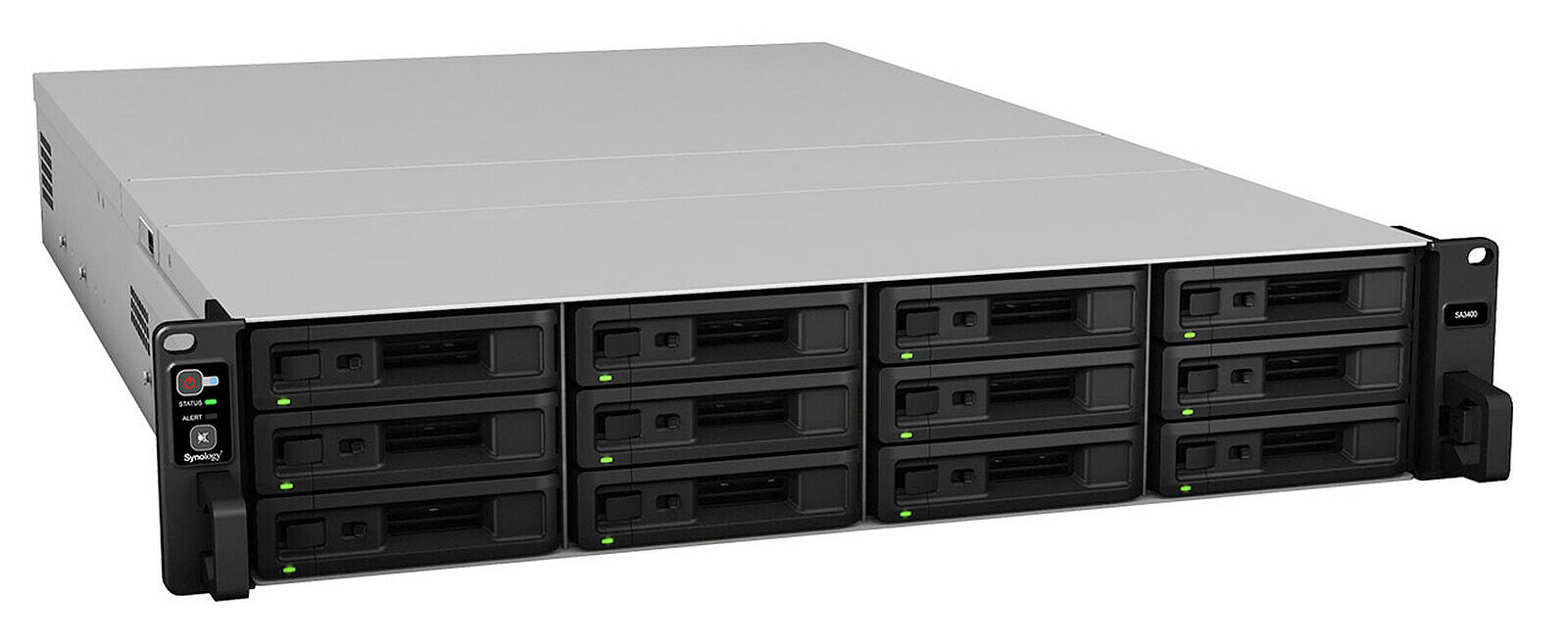
Internal NAS system
Experimental data can be stored on an internal Network Attached Storage (NAS) having a capacity of 250To (Raid 5 data redundancy system) which are seconded by two additional NAS of 100To that mirror each a part of the main NAS. This limits the risk of data loss since it can support that one of the hard drives fail without compromising the integrity of the data. The main NAS is backed up every night to the secondary NAS.
The purpose of NAS is to store and centralize data storage for local networks. The act of file serving allows for computer data to be stored on the NAS and to be easily accessible from any where within the local network. This allows for computer data to be quickly disseminated amongst its users and to share data between various computers of the lab. This NAS is accessible only from the lab and from the university building of Bichat. As such, it is not accessible from the hospital. It is possible to connect from outside (from home for instance) through VPN secured tunnel connections. This requires a specific registration through the INSERM IT service (ask Tony for the exact procedure).
Main NAS: Synology SA3400 with the RX1217sas extension module (130To)
Secondary NAS#1: Synology DS1813+ with two DX513 extension modules (107To)
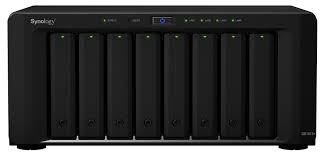
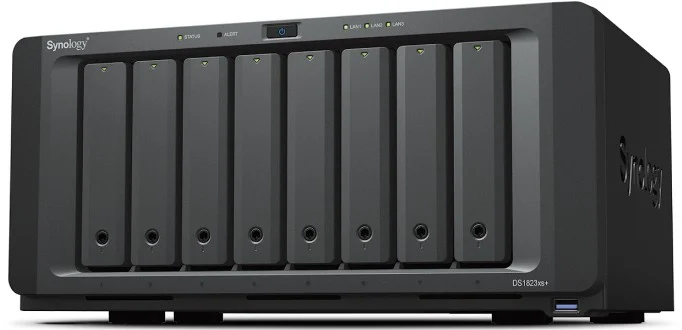
Secondary NAS#2: Synology DS1823xs+ (120To)
Important notes
• While the NAS is secured with redundant hard-drives, we cannot be certain that it will never fail, be damaged or stolen. Backup your key data on another support.
• The capacity is large but this should not prevent you from cleaning your respective working spaces.
• The password to access the NAS has been provided to your Team leader that will provide it to you upon request.
• Team-specific storage spaces are shared by all members of the Team. This means that all members from a given team have full access — write | read | erase — to the data of the other members of the team.
• The ‘Commun’ working space is not a garbage storage space. Please keep it clean.
CATI BioMéd: Bichat site shared Network Attached Storage (NAS)
This is another solution to store your data that has several important features:
• You can access it from the INSERM building but also from the University Bichat building
• You must be individually declared to use it. Contact Tony for that.
• You have access to a OwnCloud solution to synchronize a folder of your computer at work with a computer at home or when you are on the road.
You must be part of the LVTS to have access to more information…
Fab Lab
We aim at developing a ‘fab lab’ for different applications and we have already acquired several 3D printers:
Formlabs 3L / Formlab Cure L / Formlab Wash L: SLA 3D printing and post-processing (UV polymerization, Washing unit).


Ultimaker S3: dual extrusion 3D printer that delivers high-quality, composite-ready performance – in a smaller footprint.

Tenlog TL-D3 Pro Independent Dual Extruder 3D Printer. It has double nozzles that are independent of each other, and it can print large models.
