Engineered micro-structured biomimetic material for modelling the outer blood-retinal barrier
Chloé Dujardin, Walter Habeler, Paola Aprile, Alessandra Dellaquila, Christelle Monville, Didier Letourneur, Teresa Simon-Yarza
2025 • SCIENCE DIRECT
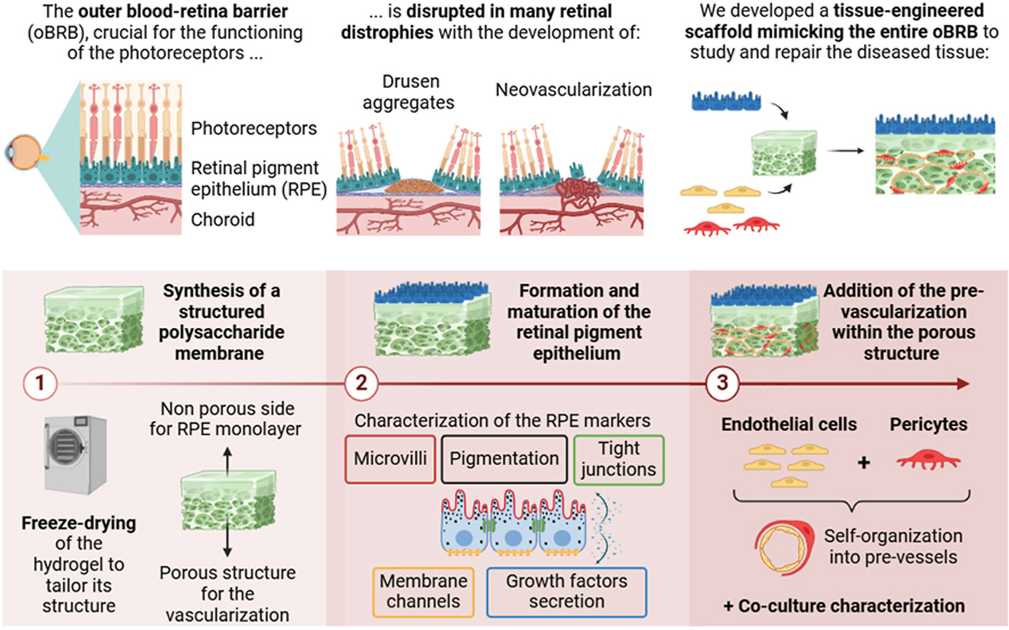
The outer blood-retinal barrier (oBRB) is compromised in several retinal pathologies, such as age-related macular degeneration affecting over 200 million people worldwide. This 200–350 μm thick tissue includes the retinal pigment epithelium (RPE), the Bruch’s membrane, and the vascularized choroid supplying the outer retina. Degeneration of the RPE and/or choroid leads to photoreceptor loss and, ultimately, blindness. Current in vitro co-culture oBRB models developed to better understand the diseases and to propose therapeutic alternatives are often simplistic, focusing on 2D cultures, or face limitations including non-physiological dimensions or low throughput.
This study presents an innovative scaffold-driven approach to model the oBRB using a polysaccharide membrane engineered by freeze-drying. Our specific protocol allowed to mimic the oBRB structure, within physiological dimensions, generating a non-porous surface to culture the hiPSC-derived RPE monolayer, and an internal 3D porous structure for the choroidal network. Results showed that the inner porous structure promoted physiological self-organization of endothelial cells and pericytes. Our single-piece functional material allowed the cultivation of both RPE and choroidal compartments in close proximity, favoring cellular interactions, while maintaining them in their designated locations. This cyto-compatible, easy-to-use, and off-the-shelf membrane, produced at large amounts and low costs, provides a physiologically relevant biomaterial for oBRB tissue modelling.